New retinal imaging technologies are helping doctors to diagnose and treat retinal diseases earlier and more effectively. These new technologies are breaking down barriers and providing hope for people with retinal diseases. In this informative blog, we will learn about three of these cutting-edge technologies and how they are changing the way we treat retinal diseases.
Introduction
The field of retinal imaging has witnessed remarkable advancements in recent years, revolutionizing our understanding of ocular health and paving the way for more accurate diagnoses and personalized treatments. The complex and intricate human eye holds valuable insights into various systemic diseases and ocular conditions.
Through the use of cutting-edge retinal imaging technologies, researchers and medical professionals can delve deeper into the intricacies of the eye and gain a comprehensive understanding of its structure, function, and pathology.
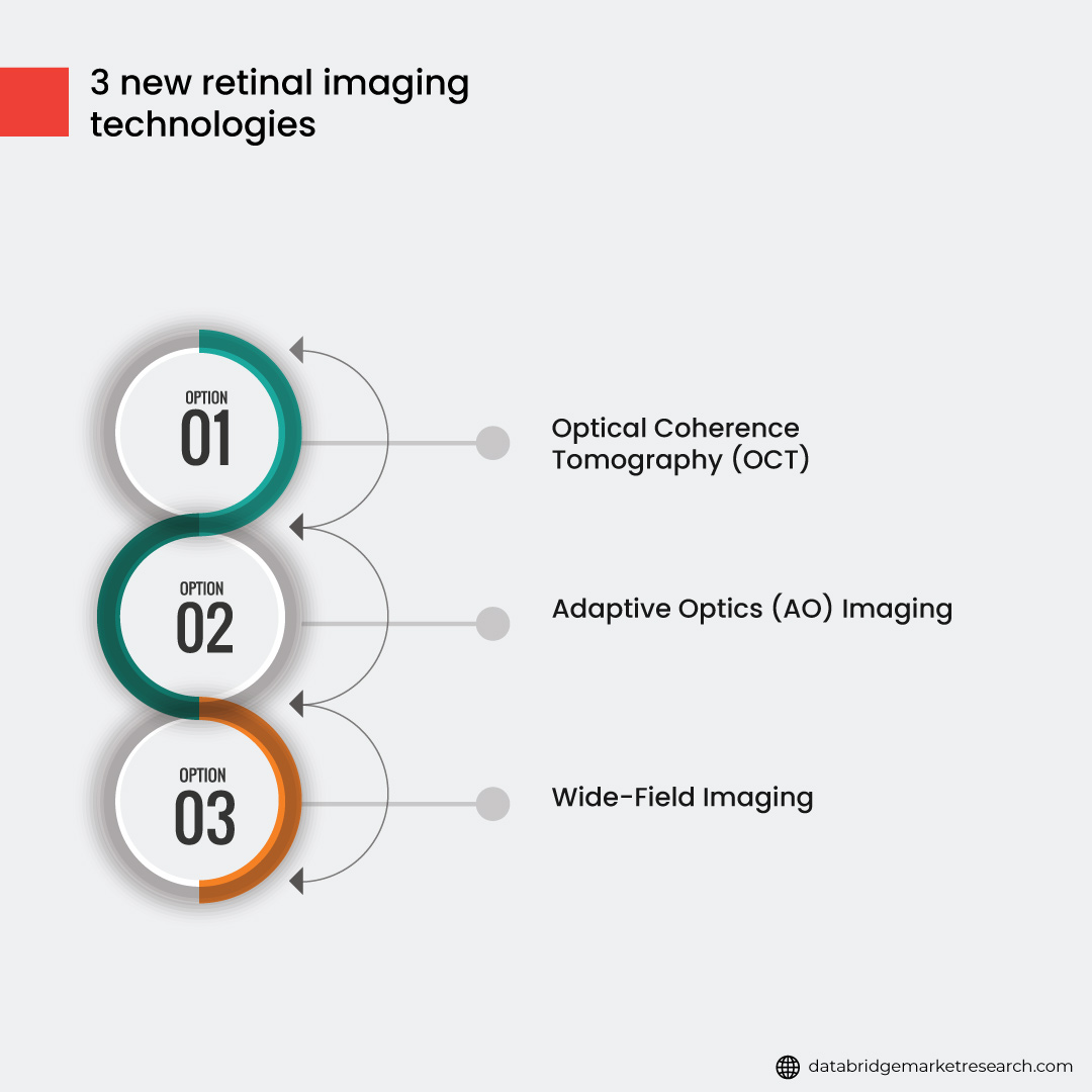
In this informative blog, we will explore the fascinating world of retinal imaging and highlight three breakthrough technologies shaping the future of ophthalmology. These following three new technologies are helping doctors to diagnose and treat retinal diseases earlier and more effectively.
Fig.1: 3 new retinal imaging technologies
- Optical Coherence Tomography (OCT)
Optical Coherence Tomography (OCT) is a non-invasive imaging technique that has revolutionized the field of ophthalmology. It provides high-resolution, cross-sectional retina images, allowing clinicians to visualize and analyze its microstructural details with exceptional precision. Let's explore each aspect of OCT in detail:
In the forecast period of 2022 to 2029, the optical coherence tomography (OCT) market is estimated to develop at a pace of 4.4%. The Data Bridge Market Research's research on the optical coherence tomography (OCT) market analyses and gives insights into the numerous aspects that are likely to be prominent during the forecast period and their influence on market growth.
To learn more about the study, visit:https://www.databridgemarketresearch.com/reports/global-optical-coherence-tomography-market
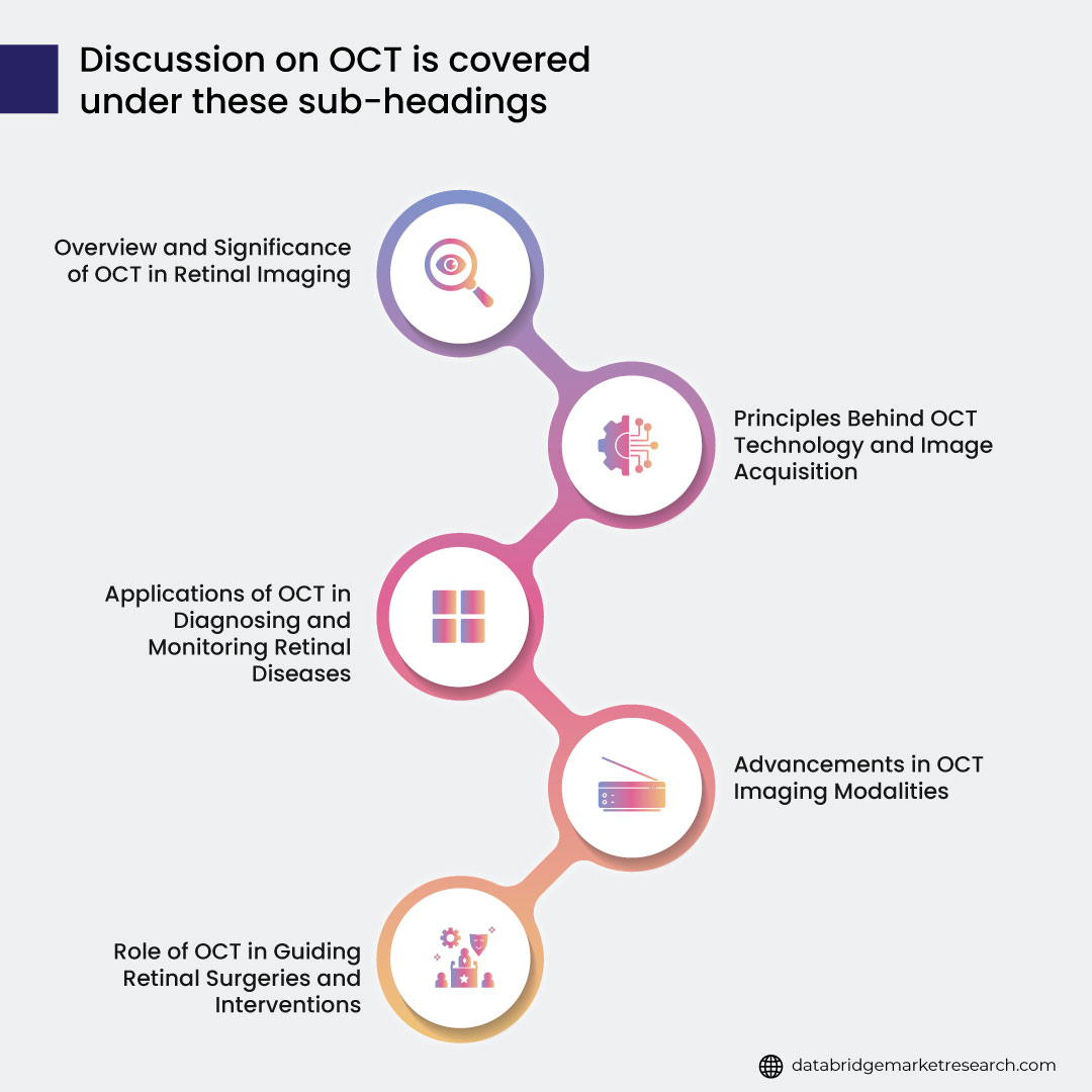
Fig.2: Discussion on OCT is covered under these sub-headings
Overview and Significance of OCT in Retinal Imaging
OCT is based on the principle of low-coherence interferometry. It uses light waves to create detailed retina images by measuring backscattered light's echo time delay and intensity. This information is then processed to generate cross-sectional and three-dimensional images of the retina.
The significance of OCT lies in its ability to provide detailed structural information about the retina, allowing clinicians to detect and monitor various retinal diseases. It enables early diagnosis, disease progression assessment, and treatment efficacy evaluation. OCT has become invaluable in managing conditions such as age-related macular degeneration (AMD), diabetic retinopathy, glaucoma, and macular edema.
Principles Behind OCT Technology and Image Acquisition
OCT employs a low-coherence light source, typically near-infrared light, which is split into a reference arm and a sample arm. The reference arm directs light to a reference mirror, while the sample arm directs light onto the retina. The backscattered light from the retina is combined with the reference light, and interference patterns are detected.
OCT creates a depth profile of the retina by measuring the intensity and time delay of the interference patterns. A series of A-scans (depth profiles) are performed across the retina to generate a cross-sectional image, and multiple cross-sectional images are combined to form a three-dimensional representation.
Applications of OCT in Diagnosing and Monitoring Retinal Diseases
OCT has become an essential tool in diagnosing and monitoring various retinal diseases. It allows clinicians to visualize and quantify structural changes in the retina, providing valuable insights into disease pathology. Some key applications include:
- AMD: OCT helps in detecting drusen (small yellow deposits), geographic atrophy (thinning of the retina), and choroidal neovascularization (abnormal blood vessel growth).
- Diabetic Retinopathy: OCT identifies macular edema, retinal thickening, and changes in the retinal layers, facilitating early intervention and treatment.
- Glaucoma: OCT aids in assessing the thickness of the retinal nerve fiber layer, identifying optic nerve head changes, and monitoring disease progression.
- Macular Disorders: OCT is crucial in evaluating conditions like macular holes, macular edema, and epiretinal membranes.
Advancements in OCT Imaging Modalities
Over time, OCT technology has advanced, leading to improvements in imaging speed, resolution, and clinical utility. Two major advancements are swept-source OCT (SS-OCT) and spectral-domain OCT (SD-OCT):
- SS-OCT: SS-OCT uses a rapidly tunable laser as a light source, which sweeps through a range of wavelengths. It offers increased imaging speed, deeper penetration into retinal tissue, and reduced motion artifacts.
- SD-OCT: SD-OCT utilizes a spectrometer to measure the spectrum of backscattered light. It provides higher resolution, faster scanning, and enhanced image quality compared to the older time-domain OCT (TD-OCT).
These advancements have significantly improved the capabilities of OCT, enabling more accurate diagnoses and better monitoring of retinal diseases.
Role of OCT in Guiding Retinal Surgeries and Interventions
OCT plays a crucial role in guiding retinal surgeries and interventions, enhancing surgical planning and real-time visualization. It provides detailed information about retinal structures such as the macula, optic disc, and retinal layers. This information helps surgeons precisely locate and navigate the surgical instruments during procedures.
OCT assists in various retinal surgeries and interventions, including:
- Vitrectomy: During a vitrectomy, which involves the removal of the vitreous gel in the eye, OCT helps in visualizing the vitreoretinal interface, detecting epiretinal membranes, and assessing retinal traction. Surgeons can use OCT to guide the removal of abnormal tissue and ensure optimal surgical outcomes.
- Retinal Detachment Repair: OCT aids in identifying retinal breaks, assessing the extent of retinal detachment, and guiding the placement of retinal tamponade agents (such as gas or silicone oil) to secure the reattachment of the retina. It allows surgeons to monitor the retinal reattachment progress during and after the procedure.
- Macular Hole Closure: In macular hole surgeries, OCT is crucial for accurate preoperative diagnosis, measuring the size and characteristics of the hole, and evaluating postoperative hole closure. Surgeons rely on OCT to guide the placement of tissue grafts or gas tamponade to facilitate hole closure and improve visual outcomes.
By integrating OCT into surgical systems, surgeons can navigate with greater precision, reduce the risk of complications, and improve surgical outcomes. Real-time OCT feedback enables the immediate assessment of tissue changes, making surgeries more efficient and enhancing patient safety.
- Adaptive Optics (AO) Imaging
Adaptive Optics (AO) imaging is an innovative technology that overcomes the limitations of conventional retinal imaging techniques, allowing for high-resolution imaging of the retina at the cellular level. Let's explore each aspect of AO imaging in detail:
The laser guide star adaptive optics market will reach an estimated value of USD 2,808.65 million and grow at a CAGR of 30.10% from 2021 to 2028.
To know more about the study, visit: https://www.databridgemarketresearch.com/reports/global-laser-guide-star-adaptive-optics-market
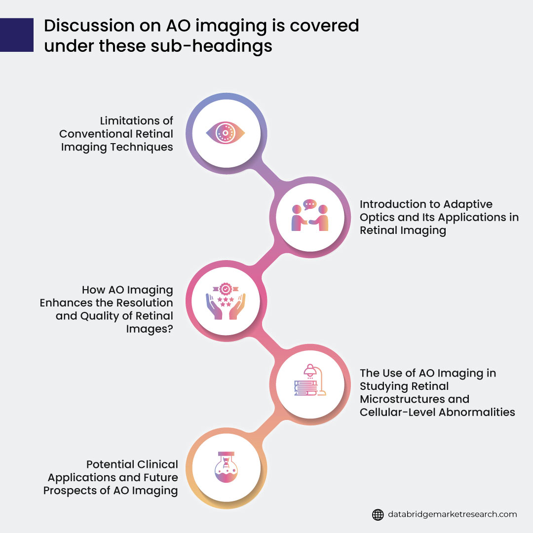
Fig.3: Discussion on AO imaging is covered under these sub-headings
Limitations of Conventional Retinal Imaging Techniques
Conventional retinal imaging techniques, such as fundus photography and scanning laser ophthalmoscopy, have resolution and image quality limitations. These techniques are affected by various factors, including optical aberrations of the eye and scattering of light within the eye's media. As a result, the images obtained lack fine details and clarity, making it challenging to visualize microscopic structures and cellular abnormalities in the retina.
Introduction to Adaptive Optics and its Applications in Retinal Imaging
Adaptive optics was originally developed for astronomy to correct atmospheric distortions in telescope imaging. It has been adapted for retinal imaging to correct optical aberrations in the human eye. AO systems utilize a wavefront sensor to measure the aberrations and deformable mirrors to correct for these aberrations in real-time dynamically. This correction enables high-resolution imaging of the retina.
- In retinal imaging, AO systems are used to obtain detailed images of retinal microstructures, such as photoreceptor cells, retinal pigment epithelium, and retinal blood vessels. AO imaging also allows visualization of cellular-level abnormalities, including microaneurysms, drusen, and individual retinal nerve fiber bundles. This level of detail provides valuable insights into retinal pathologies and helps in early detection and monitoring of diseases.
How AO Imaging Enhances the Resolution and Quality of Retinal Images?
AO imaging improves the resolution and quality of retinal images by correcting for optical aberrations specific to an individual's eye. The deformable mirrors in AO systems dynamically adjust to counteract the distortions caused by the eye's optical system. This correction results in sharper images with improved spatial resolution, allowing for better visualization of retinal structures.
By reducing the impact of aberrations, AO imaging can capture details at the cellular level, revealing structures that are not discernible with conventional imaging techniques. This enhanced resolution and image quality enable clinicians and researchers to study the retina in greater detail and accurately assess retinal health.
The Use of AO Imaging in Studying Retinal Microstructures and Cellular-Level Abnormalities
AO imaging provides a unique opportunity to study retinal microstructures and cellular-level abnormalities. Researchers can investigate photoreceptor cells' morphology, density, and arrangement, which play a critical role in vision. It allows for assessing changes in these cells over time and in response to various retinal diseases.
AO imaging also aids in the identification and characterization of cellular-level abnormalities associated with retinal pathologies. For example, it helps detect and monitor retinal microvascular changes in diabetic retinopathy or the presence of individual drusen in age-related macular degeneration.
Potential Clinical Applications and Future Prospects of AO Imaging
AO imaging holds tremendous potential in clinical applications and research. Some potential areas of application include:
- Early Diagnosis and Monitoring of Retinal Diseases: AO imaging can provide early detection and precise monitoring of retinal pathologies, allowing for timely intervention and personalized treatment plans.
- Evaluation of Treatment Efficacy: AO imaging can be used to assess the effectiveness of therapeutic interventions, such as anti-vascular endothelial growth factor (anti-VEGF) therapy in neovascular retinal diseases.
- Customized Treatment Planning: AO imaging can aid in designing personalized treatment strategies based on individual retinal characteristics and disease progression.
- Advancing Our Understanding of Retinal Physiology and Disease Mechanisms: AO imaging enables researchers to study the retina's normal and abnormal cellular architecture and how it relates to visual function. This deeper understanding can lead to the developing new therapeutic targets and interventions.
The future prospects of AO imaging are promising. Here are some potential advancements and applications:
- Integration with Other Imaging Modalities: Combining AO imaging with other imaging techniques, such as OCT or fluorescein angiography, can comprehensively assess retinal structure and function. This integration could offer a more holistic approach to the diagnosis and monitoring of retinal diseases.
- Treatment Guidance in Real-Time: AO imaging has the potential to be used intraoperatively to guide surgical procedures, such as retinal gene therapy or implantation of retinal prostheses. Real-time feedback from AO imaging can enhance surgical precision and improve outcomes.
- Monitoring Treatment Response and Disease Progression: AO imaging can be used longitudinally to track disease progression and assess treatment response. It may help clinicians evaluate the efficacy of novel therapies and facilitate personalized treatment plans.
- Early Detection of Neurodegenerative Diseases: Research suggests that changes in the retinal microstructure may precede the onset of neurodegenerative diseases, such as Alzheimer's and Parkinson's. AO imaging could serve as a non-invasive tool for early detection and monitoring of these conditions.
- Telemedicine and Remote Monitoring: AO imaging systems are becoming more compact and portable, making them potentially suitable for telemedicine applications. Remote imaging and monitoring of retinal structures using AO imaging could enhance accessibility to high-quality retinal care, especially in underserved areas.
- Artificial Intelligence Integration: Combining AO imaging with artificial intelligence algorithms could automate image analysis and aid in the detection and classification of retinal abnormalities. This integration could improve efficiency, accuracy, and scalability in clinical practice.
In summary, adaptive optics (AO) imaging offers exciting potential for advancing the field of retinal imaging. Its ability to enhance resolution, visualize retinal microstructures, and detect cellular-level abnormalities opens up new possibilities for early diagnosis, personalized treatment, and our understanding of retinal diseases. With further advancements and integration with other technologies, AO imaging is poised to play a vital role in shaping the future of ophthalmology and improving patient care.
- Wide-Field Imaging
Traditional fundus photography has limitations in capturing a comprehensive view of the retina. It provides a limited field of view, typically capturing only the central region of the retina. This restricts the ability to detect and assess peripheral retinal pathology, which can be crucial in diagnosing and managing various retinal diseases. Wide-field imaging technologies have emerged as a solution to overcome these limitations. Let's delve into the details of wide-field imaging:
Data Bridge Market Research analyses that the wide field imaging devices market which was USD 531.28 million in 2021, would rocket up to USD 926.58 million by 2029, and is expected to undergo a CAGR of 7.20% during the forecast period 2022 to 2029.
To know more about the study, visit: https://www.databridgemarketresearch.com/reports/global-wide-field-imaging-devices-market
Fig.4: Discussion on wide-field imaging is covered under these sub-headings
Limitations of Traditional Fundus Photography
Traditional fundus photography captures a small portion of the retina, usually limited to the macula and optic nerve head. This restricted field of view can lead to missed peripheral retinal pathology, such as peripheral lesions, tears, or detachments. These peripheral abnormalities are significant in conditions like diabetic retinopathy, retinal vascular occlusions, and peripheral retinal degeneration. Therefore, a broader view of the retina is necessary to evaluate and manage retinal diseases comprehensively.
Introduction to Wide-Field Retinal Imaging Technologies
Wide-field retinal imaging technologies provide a more extensive view field than traditional fundus photography. They encompass panoramic imaging techniques that enable visualization of a broader area of the retina, including the peripheral regions. Wide-field imaging systems employ specialized optics and sensors to capture and generate detailed images of the retina.
Advantages of Wide-Field Imaging in Detecting Peripheral Retinal Pathology
Wide-field imaging offers several advantages in the detection and evaluation of peripheral retinal pathology:
- Enhanced Visualization: Wide-field imaging provides a more comprehensive and detailed view of the retina, enabling the identification of peripheral lesions, retinal tears, or detachments that may have clinical significance.
- Early Detection: Peripheral retinal pathology, including retinal breaks, lattice degeneration, and peripheral lesions, can be detected earlier with wide-field imaging. This early detection allows for prompt intervention and preventive measures to avoid complications.
- Treatment Planning and Monitoring: Wide-field imaging aids in treatment planning by identifying the extent of peripheral retinal pathology. It facilitates accurate targeting of laser photocoagulation, cryotherapy, or surgical intervention, especially in conditions like retinopathy of prematurity or retinal detachments. Wide-field imaging also enables post-treatment monitoring to assess treatment response and identify any new pathology.
Clinical Applications and Benefits of Wide-Field Imaging In Various Retinal Diseases
Wide-field imaging has found utility in several retinal diseases:
- Diabetic Retinopathy: Wide-field imaging helps in detecting peripheral ischemia, non-perfusion areas, and neovascularization beyond the posterior pole. This aids in determining the severity of diabetic retinopathy and guiding appropriate treatment, such as pan-retinal photocoagulation.
- Retinal Vascular Occlusions: Wide-field imaging allows for a comprehensive assessment of retinal vascular occlusions, including central retinal vein occlusion (CRVO) and branch retinal vein occlusion (BRVO). It helps in identifying areas of non-perfusion, neovascularization, and retinal ischemia beyond the posterior pole, guiding treatment decisions such as anti-vascular endothelial growth factor (anti-VEGF) therapy or laser photocoagulation.
- Retinopathy of Prematurity (ROP): Wide-field imaging plays a crucial role in the screening and monitoring of ROP, a potentially blinding condition affecting premature infants. It helps identify the extent and location of the disease, enabling timely intervention with laser therapy or anti-VEGF injections to prevent progression to retinal detachment.
- Retinal Degenerations: Inherited retinal degenerations, such as retinitis pigmentosa, can exhibit peripheral retinal abnormalities. Wide-field imaging allows for the evaluation of the extent and characteristics of degenerative changes in the peripheral retina, aiding in disease staging, prognosis determination, and potential treatment considerations.
- Retinal Tears and Detachments: Wide-field imaging is valuable in diagnosing and managing retinal tears and detachments. It helps identify peripheral retinal breaks and their association with vitreoretinal traction, facilitating appropriate treatment strategies such as laser photocoagulation, cryotherapy, or vitreoretinal surgery.
- Uveitis: Wide-field imaging assists in evaluating the extent of the inflammation and associated complications in uveitis, including peripheral involvement. It aids in monitoring disease progression, assessing treatment response, and guiding targeted interventions such as immunosuppressive therapy or intraocular injections.
- Choroidal Neovascularization (CNV): Wide-field imaging allows for detecting and monitoring CNV, particularly in conditions like age-related macular degeneration (AMD). It helps visualize the extent of CNV beyond the macula, guiding treatment decisions such as anti-VEGF therapy or photodynamic therapy.
- Optic Nerve Disorders: Wide-field imaging can provide valuable information about the optic nerve head and its surrounding structures. It aids in the assessment of optic nerve head drusen, optic disc edema, and other optic nerve pathologies, allowing for early detection and management.
The benefits of wide-field imaging in these retinal diseases include improved diagnostic accuracy, enhanced monitoring of disease progression, and precise treatment planning. It offers a more comprehensive evaluation of the retina, especially in the periphery, leading to better management and improved visual outcomes for patients. Wide-field imaging has become an indispensable tool in the armamentarium of ophthalmologists, enabling them to provide comprehensive care for a wide range of retinal conditions.
In Summary
In conclusion, the field of retinal imaging has witnessed remarkable advancements in recent years, breaking barriers and revolutionizing how we visualize and understand the intricacies of the retina. Cutting-edge technologies such as Optical Coherence Tomography (OCT), Adaptive Optics (AO) imaging, and Wide-Field Imaging have emerged as powerful tools, each offering unique insights and capabilities in retinal imaging.
OCT has proven to be a game-changer in diagnosing and monitoring retinal diseases. Its non-invasive nature, high-resolution imaging, and ability to visualize retinal layers have transformed clinical practice. From the principles behind OCT technology to its applications in guiding retinal surgeries and interventions, OCT continues to pave the way for better patient care.
Adaptive Optics imaging has revolutionized our ability to visualize retinal microstructures and cellular-level abnormalities. By overcoming the limitations of conventional imaging techniques, AO imaging enhances the resolution and quality of retinal images, providing valuable insights into retinal physiology and disease mechanisms. Its potential clinical applications, including customized treatment planning and early detection of neurodegenerative diseases, hold great promise for the future.
Wide-Field Imaging has expanded our perspective by capturing a broader view of the retina, including the peripheral regions. This panoramic imaging approach has proven invaluable in detecting peripheral retinal pathology and guiding treatment decisions in various retinal diseases. Its benefits in conditions such as diabetic retinopathy, retinal vascular occlusions, and retinopathy of prematurity cannot be overstated.
The combination of these cutting-edge retinal imaging technologies is transforming the field of ophthalmology. They have provided clinicians unprecedented insights into retinal health and disease, enabling earlier detection, personalized treatment approaches, and improved patient outcomes.
As technology continues to evolve, we can anticipate further advancements in retinal imaging. Integration with artificial intelligence, telemedicine applications, and continued refinements in imaging modalities hold immense potential for the future. These developments will enhance diagnostic accuracy, more effective treatment strategies, and a deeper understanding of retinal diseases.
In conclusion, the exploration of cutting-edge retinal imaging technologies is breaking barriers and opening new frontiers in the field of ophthalmology. Through these innovative techniques, we are unraveling the mysteries of the retina and improving the lives of countless individuals affected by retinal diseases. The journey of discovery in retinal imaging continues, and the future holds even more exciting possibilities.


