Oral cancer and head and neck cancer are among the top 10 most prevalent types of cancer. Approximately 250 people in New Zealand are diagnosed with mouth cancer each year. While most instances will be effectively treated, oral cancer must be diagnosed early to increase the chances of success. Early detection is critical since oral cancer therapy can significantly impact someone's life. When patients are diagnosed with oral cancer in its later stages and receive therapy, their daily lives can become extremely challenging since oral cancer treatment affects how they talk, chew, swallow, and engage socially.
VELscope, also known as the VELscope Vx Enhanced Oral Assessment System, is a wireless handheld device that employs natural tissue fluorescence to improve our capacity to detect oral abnormalities that are typically invisible to the human eye. It is frequently utilized as an adjunctive tool for imaging tissue abnormalities in the mouth since it is convenient, easy to use, and employs basic and straightforward technology.
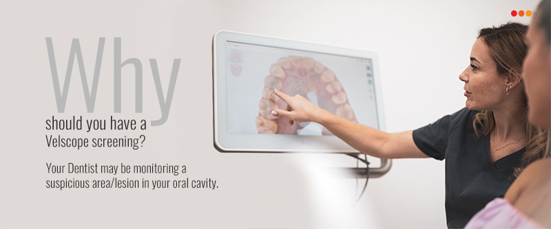
VELscope is a particular type of light to detect abnormalities, effectively used to see early forms of cancer or disease during an oral cancer screening. The VELscope, according to Gabriela Videira, MD of ISCS-Sul and Mário Arajo, RDH, BS, does not detect whether or not an oral lesion is malignant. Instead, the technology just aids in detecting anomalies that are not evident to the human eye.
Data Bridge Market Research analyses that the oral cancer treatment market, USD 1,805.8 million in 2022, would rise to USD 2,729.72 million by 2030 and is expected to undergo a CAGR of 5.30% during the forecast period 2023 to 2030. The oral cancer treatment market is segmented on the basis of type, therapy type, treatment, route of administration, end users, and distribution channel. North America dominates the market in the forecast to the rising affordability of drugs and the surge in the cases of oral cancer. Furthermore, growing awareness among the people in this region to cure dental caries and technological developments due to the presence of major market players lead to market growth.
To learn more about the study, visit: https://www.databridgemarketresearch.com/reports/global-oral-cancer-treatment-market
How Does VELscope Work?
Unlike traditional oral cancer screening, which includes visually inspecting the mouth for red, white, or black spots, VELscope employs a superior blue light to stimulate molecules deep inside the layer of our oral mucosal tissues, such as the mucous membrane lining the interior of the mouth. These energized molecules then emit their own light in green, yellow, and red wavelengths. The VELscope's unique filter enables fluorescence imaging by suppressing reflected blue light and increasing the contrast between normal and diseased tissue. Essentially, VELscope enables dentists to detect dysplastic cells or cells that are just starting to change into pre-cancerous cells via aberrant fluorescence patterns. A variety of factors can contribute to abnormal fluorescence patterns, including:
- Metabolic activity in epithelial tissue has increased
- The fluorescent collagen cross-links in the connective tissue layer underlying the basement membrane were broken down
- An increase in tissue blood content is caused by inflammation or angiogenesis because hemoglobin absorbs fluorescence excitation (blue light) strongly
- Melanin or amalgam particles that absorb light are present.
Oral abnormalities are not visible to the naked eye can now be discovered using VELscope. Furthermore, doctors may now determine the proper margins for surgical excision.
It only takes two or three minutes to complete the VELscope inspection. Every single year, many lives are saved because of this non-invasive, painless surgery. A quick description of a VELscope inspection is provided below:
The dentist will first conduct a standard visual examination of the entire lower face. Along with the teeth, this also refers to the glands, tongue, cheeks, and palate. After that, the mouth is rinsed with a pre-rinse solution for under a minute. The dentist gives patients protective eyewear to safeguard the retinas' structural integrity. To clearly see the oral cavity, the room's lights are decreased.
Oral Cancer and VELscope
The most frequent cause of mouth cancer growth is frequent smoking. Another element that has been linked to a higher risk of mouth cancer is alcohol consumption. On the other side, exposure to the sun may raise your risk of developing lip cancer. You can lower your risk of acquiring mouth cancer by giving up smoking, abstaining from alcohol, using lip balm and sunscreen every day, and quitting drinking.
You naturally have an increased risk of having mouth cancer if you are above forty. Oral cancer risk may also increase as a result of genetic factors. Men are twice as likely as women to have oral cancer. You must go to dental exams and screenings because none of these risk factors are under our control. The most frequent cause of mouth cancer growth is frequent smoking. Alcohol consumption can also raise your risk. Additionally, exposure to the sun may increase your risk of developing lip cancer. You can reduce your chance of mouth cancer by giving up smoking, abstaining from drinking, and using sunscreen.
Since it claims more lives each year than combined testicular, cervical, and brain cancer but receives less attention, oral cancer is frequently referred to as the "forgotten disease." Only 57% of the more than 30,000 Americans who develop oral cancer each year will survive for more than five years without treatment.
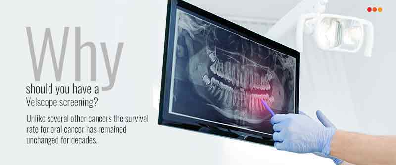
Many people think that if they don't use alcohol or tobacco, they won't get mouth cancer. Alcohol and tobacco use does increase the risk of oral cancer, yet 25% of people with the disease abstain from both. Getting annual oral cancer screenings is the greatest method to prevent mouth cancer. Most dentists examine for oral cancer as part of a routine dental examination. The FDA-approved VELscope gives dentists another exam tool to aid in the early detection of oral cancer. The VELscope is a blue excitation lamp that draws attention to alterations in malignant and pre-cancerous cells.
The Velscope System
Let's look at the VELscope itself to understand better how it functions. The VELscope depends on two essential parts: an eyepiece with an inbuilt optical filter and an LED ring that emits a specific wavelength of blue light. Clinicians can see areas that need more research by observing cellular and structural tissue changes while beaming light into the oral cavity and looking at the patient's oral mucosa through an optical filter. We now arrive at the concept of tissue fluorescence.
Tissue Reflectance VS Tissue Fluorescence
Utilizing the principle of tissue reflectance and conventional white light, doctors conduct oral examinations using traditional oral mucosal examination equipment. In contrast, the VELscope uses the idea of tissue fluorescence rather than reflectance. Fluorophores, which are chemical substances that respond to light excitation, are what produce tissue fluorescence. Fluorophores respond to the blue light of the VELscope by generating their own light at a longer wavelength, which can be seen through the VELscope eyepiece's optical filter. The VELscope can see the cellular change in the oral mucosa due to the fluorophores' stimulation, or lack thereof in some cases.
Fluorescence Patterns
The fluorophores in the oral mucosa's tissues normally respond to blue light when observed with the VELscope, as evidenced by the brilliant apple-green color of normal fluorescence patterns. Lymphoid clusters, the fungiform papillae on the tongue, and the extensively vascularized anterior tonsillar pillars are excellent illustrations of how normal fluorescence patterns can also reveal a lack of fluorescence. During a standard white-light reflectance assessment, sick mucosal regions may go unnoticed by the human eye, but abnormal fluorescence patterns enable clinicians to spot them. Normal causes of abnormal fluorescence patterns include:
- An increase in the epithelium's metabolic activity
- Fluorescent collagen cross-links in the connective tissue layer below the basement membrane are broken down
- An increase in tissue blood volume caused by angiogenesis or inflammation (hemoglobin absorbs fluorescence excitation light (blue) and emission light (green) very strongly)
- The presence of light-absorbing pigments, such as melanin or amalgam particles
Although the VELscope is frequently referred to as an instrument for screening for oral cancer, the idea of tissue fluorescence encompasses a wide range of oral abnormalities, enabling the VELscope to assist in visualizing a variety of oral health issues:
- Bacterial, fungi, and viral infections
- Several sources of inflammation, such as lichen planus and other lichenoid reactions
- Trauma
- Squamous papilloma's and salivary gland tumors are examples of oral cancer
- Tooth dysplasia
Data Bridge Market Research analyses that the North America oral care products and other dental consumables market will exhibit a CAGR of around 2.99% for the forecast period of 2021-2028. The growing prevalence of dental disorders, increased focus on research and development proficiencies in regards to medical devices and on the adoption of advanced healthcare technologies, ever-rising geriatric population and rising expenditure on the healthcare infrastructure development especially in the developing economies are the major factors attributable to the growth of oral care products and other dental consumables market. The North America oral care products and other dental consumables market is segmented on the basis of product type and distribution channel.
To learn more about the study, visit: https://www.databridgemarketresearch.com/reports/north-america-oral-care-products-other-dental-consumables-market
Conclusion
An innovative handheld tool called VELscope offers hygienists and dentists an easy-to-use supplementary mucosal examination system for the early identification of aberrant tissue. A harmless blue light is emitted into the oral cavity via the VELscope hand piece, which excites the tissue and causes it to fluoresce from the epithelium's surface down to the basement membrane (where premalignant alterations generally start) and into the stroma below. Normal tissue typically glows brightly apple-green, but a lack of fluorescence distinguishes suspicious areas and hence seem dark.

According to reports and clinical experience, VELscope technology is useful for further analyzing oral health conditions. Clinicians also claim that incorporating technology into dentistry is quite simple because it can readily photograph any questionable spots for possible surgical biopsies. VELscope has been recognized by the World Health Organization's Department of Essential Health Technologies as a novel medical technology that addresses global health challenges and is "likely to be available, appropriate, and affordable for use in low- and middle-income countries." This demonstrates that the VELscope is a necessary tool for improving oral health in a cost-effective and user-friendly manner.
Fluorescence Visualization (FV) is used in an innovative new method by the VELscope. In essence, abnormalities and lesions that are normally unseen to the naked eye are exposed in the mouth by shining intense blue light into it. The fact that oral cancer symptoms might be mistaken for those of less serious conditions makes diagnosis of the disease one of the most challenging tasks. The VELscope System gives the dentist valuable information about what is going on below the surface.
The healthy soft tissue of the mouth naturally absorbs the VELscope frequency of blue light. Healthy soft tissue beneath the skin appears green, and the problem areas turn much darker. The following are some benefits of utilizing the VELscope system:
- Combination with digital photography is possible
- Detects white and red spots, as well as lesions
- Detects trouble spots that are invisible in white light
- Exposes tissue that is malignant and pre-cancerous
- FDA-approved
- Aids dentists in making sure all unhealthy soft tissue has been removed
- Increases the chance of survival by assisting in the early diagnosis of oral cancer
- Swift and painless inspections
Global cosmetic dentistry market was valued at USD 23.45 billion in 2021 and is expected to reach USD 40.60 billion by 2029, registering a CAGR of 7.10% during the forecast period of 2022-2029. The "dental hospitals and clinics" account for the largest end user segment in the cosmetic dentistry market within the forecasted period as they provide the government recognized dentists. The market report curated by the Data Bridge Market Research team includes in-depth expert analysis, patient epidemiology, pipeline analysis, pricing analysis, and regulatory framework. The cosmetic dentistry market is segmented on the basis of product, age group and end user.
To learn more about the study, visit: https://www.databridgemarketresearch.com/reports/global-cosmetic-dentistry-market