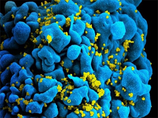
Scientists from EMBL Heidelberg and the Center for Infectious Diseases at Heidelberg University Hospital have for the first time succeeded in mapping HIV during transport into the nucleus of an infected cell. The scientists have found that the virus penetrates the nuclear pores (the openings in the membrane around the cell nucleus through which molecules can enter and exit) intact and only breaks in the nucleus, where it releases its genetic information. This illustrates an important mechanism by which the genetic material of the virus is integrated into the genome of the infected cell. The human immunodeficiency virus type 1 (HIV-1), which is the focus of this study, primarily infects certain cells of the immune system and thereby severely weakens the body's defense against diseases is securely packaged in a cone-shaped protein capsule, the so-called capsid, which consists of individual hexagonal parts.
Scientists knew the mechanism of capsid but not how the genetic material is transferred from the capsid to the cell nucleus, where the formation of new viruses is triggered. Work of Heidelberg suggests using newly developed methods for 3D imaging of molecular complexes in virus-infected cells; the scientists were able to image the virus capsid directly during transport to the cell nucleus. So far, the capsid shouldn't penetrate through the pores. However, the question of how the viral genome gets into the cell nucleus is essential for its reproduction. Therefore, our results support the search for new targets for future therapeutic approaches. Although current treatment options can suppress the replication of the virus in the body, a real cure that eliminates the virus is not yet possible.
In order to get a detailed insight into the inner workings of infected immune cells in the laboratory, the scientists used high-resolution imaging processes. With the help of the central electron microscopy facility of Heidelberg University and the cryo electron microscopy service platform of EMBL Heidelberg, they combined optical and electron microscopic methods and were able to reconstruct 3D images of molecular structures from their data. It additionally, allowed them to visualize the composition and architecture of viral complexes and their interaction with cellular structures in high resolution.