Around 20 million individuals are newly diagnosed with cancer each year, and nearly 10 million people pass away. While malfunctioning proteins are the primary causes of malignancies that disturb cellular homeostasis, the relationship between genetic abnormalities and cancer is well-recognized in the field of cancer research. Until now, cancer prevention has focused on reducing the negative consequences of gene alterations. However, there is an increasing trend in chemoprevention to alter the proteins that are encoded by these damaged genes. For instance, the BRCA1 protein helps to repair DNA damage, which plays a role in tumor suppression. Less functional BRCA1 individuals develop increased genetic instability, which increases the risk of cancer induction.
Single particle analysis and cryo-tomography are two examples of the cryo-electron microscopy (cryo-EM) family of methods used to generate high-resolution structural data for biological systems. In cryo-EM, sample solutions are quickly frozen in order to preserve the specimens' natural structure and stop crystalline ice from forming. This causes a suspension of individual proteins orientated at various random angles within the ice, which is what is observed in single-particle analysis. The proteins are then visualized using a cryo-transmission electron microscope (cryo-TEM), creating hundreds of 2D projections of the sample. Combining these projections can then create a high-resolution 3D model of the protein. Atomic resolution is conceivable and becoming more and more available with contemporary electron detectors and analytic tools.
By combining cryo-EM and 3D reconstruction, a number of significant advances in structural biology have been realized over the past few years. Hardware and software advancements have been a major factor in cryo-EM development. The image recording device and electron microscope are examples of the hardware; 3D reconstruction techniques and image data processing are examples of the software.
Data Bridge Market Research analyses a growth rate in the global cryo-electron microscopy market in the forecast period 2022-2029. The expected CAGR of global cryo-electron microscopy market is tend to be around 8.67% in the mentioned forecast period. The market was valued at USD 725.43 million in 2021, and it would grow upto USD 1410.82 million by 2029. The global cryo-electron microscopy market is segmented on the basis of type, product type, component and application.
To know more about the study, visit: https://www.databridgemarketresearch.com/reports/global-cryo-electron-microscopy-market
The Concept in Brief
It is becoming more commonplace to use cryo-electron microscopy (cryo-EM) to examine the molecular architecture of protein assemblies, viruses, and organisms. This page focuses on several aspects of cryo-EM, including its benefits and drawbacks, applications, how it differs from EM techniques, and recent studies utilizing cryo-EM technology. The term "cryo-EM" refers to a type of electromagnetic imaging (EM) in which radiation-sensitive specimens are imaged using a transmission electron microscope (TEM) under cryogenic settings. The term "cryo-EM" is frequently used to refer to several experimental techniques, including cryo-electron tomography, electron crystallography, and single-particle cryo-EM.
Each cryo-EM technique can be utilized independently or as part of hybrid approaches that combine cryo-EM data with complementing data from X-ray crystallographic and nuclear magnetic resonance (NMR) spectroscopy approaches. For imaging biological specimens including bacteria, plunge-frozen cells, and whole tissue sections, cryo-EMs are becoming more widely available and considerably simpler to operate.
Researchers are currently using Cryo-EM to aid their work on cancer, advancing the discipline of "structural oncology," which aims to employ structural biology to combat cancer. Cryo-high-resolution EM's analytical capabilities are poised to transform the way scientists study oncology by enabling focused observation, analysis, and ultimately treatment.
EM and Cryo-EM
Staining, dehydration, and chemical fixation of specimens are used in traditional EM procedures to produce the image. In EM, the interaction of organic matter and electrons severely harms biological specimens. In contrast, since cryo-EM does not require additional imaging techniques, it preserves the specimens' original hydrated state. The electron irradiation breaks the specimens' chemical bonds, resulting in the production of free radicals that further damage the samples. Low electron doses produce noisy images even though they can help conserve specimens by reducing radiation damage.
Cryo-EM can solve this problem efficiently since it uses frozen specimens that are kept at liquid helium or liquid nitrogen temperatures for imaging, which lessens the damaging effects of electron irradiation on the specimens. The biological samples are photographed at liquid helium or liquid nitrogen temperatures after being quickly vitrified in an ice layer that resembles glass. Radiation damage is much diminished at nitrogen temperature, enabling the employment of a larger electron dose to produce images with a good signal-to-noise ratio. Through the cryo-EM technique, it is possible to obtain three-dimensional (3D) reconstructions of specimens at a near-molecular level in liquid nitrogen and helium. But how does cryo-EM work?
-
Freezing- A grid is coated with the sample, which is after that frozen in liquid ethane and kept in liquid nitrogen. It must freeze quickly enough to stop any water present from forming ice crystals. Ice lattices are likely to cause structural damage to the material, absorbing the electron beam and obscuring the image. Water solidifies as an amorphous solid (vitreous ice) and does not crystallize if the sample is frozen rapidly enough.
-
Recording- Based on the interactions of the substance with an electron beam, pictures of many copies of the molecule (or other material, such as viruses) suspended in random orientations in vitrified ice are captured. Images with more recent "direct electron detectors" are of higher quality than those taken with previous digital cameras.
-
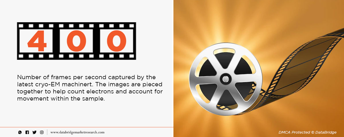
Data Processing- The sample's structure is ascertained by merging the many molecular perspectives into a 3D model. Sometimes the average of tens of thousands or even hundreds of thousands of particle images constitutes the final image, or density map. Hundreds of thousands of frames per second are captured to track the motion of the particles and prevent resolution loss.
Early cryo-EM applications were constrained by the need to employ low power electron beams to preserve the sample, resulting in low-resolution images. High resolution images may now be created with fewer electrons thanks to advancements made in direct electron detector technology. The direct electron detector is a camera that can quickly capture photos of a single molecule at dozens of frames per second by having an improved ability to detect electrons. The creation of better image-processing techniques has also facilitated the revolution in cryo-EM. Such algorithms are needed to determine picture orientation and align the images in order for methods that rebuild a three-dimensional image from two-dimensional images. Technological improvement has lowered the limit size of the samples that can be frozen, allowing individual proteins to be analyzed after imaging.
Advantages of Cryo-EM Technology
Nuclear magnetic resonance spectroscopy and X-ray crystallography were earlier structural biology methods. Due to the requirement for high sample sizes, these approaches have only had limited applicability. In order to perform X-ray crystallography, specimens must be crystallized, a challenging procedure that alters the environment to one that is non-physiological.
Cryo-EM is well suited for imaging structures at almost atomic resolution because it doesn't require large sample quantities or crystallization. The approach also allows the specimen to be analyzed in its natural physiological environment because it does not chemically fix or stain it. Furthermore, structures can be flash-frozen in a variety of conformations to enable the inference of biological systems without the restriction of crystals freezing the sample in a static pose.
The fact that the molecule of interest does not have to be crystallized is a significant advantage of cryo-EM over x-ray crystallography. Some proteins or important macromolecules simply cannot be crystallized; others have their structures irreversibly altered as a result of crystallization. Unlike x-ray crystallography, which can only determine a single structure, proteins can be seen in all of their conformations.
Cryo-EM samples, unlike conventional electron microscopy, are not dehydrated or stained, so their structure remains close to the true shape of the hydrated structure in their native environment, with no false shapes created by the stain. Freezing samples reduces the amount of radiation damage that can occur as a result of electron irradiation. Frozen samples are also less likely to be damaged by the electron microscope's low pressure/vacuum conditions.
Disadvantages of Cryo-EM Technology
Images often have a very low signal-to-noise ratio due to a lack of stain and, thus a lack of contrast, necessitating highly advanced detection hardware and image processing.
Specimen preparation can be difficult, as it is necessary to optimize ice thickness and particle distribution. Proteins can sometimes adopt preferred orientations, making 3D reconstruction impossible.
The most advanced cryo-EM equipment is still prohibitively expensive. The creation of centralized facilities may aid in increasing access to cryo-EM equipment. Cryo-EM, on the other hand, is ineffective for imaging very small proteins and takes a long time to generate sample images. Furthermore, the technique necessitates very high sample homogeneity, which complicates obtaining high-resolution images of flexible proteins. Furthermore, the current resolution of cryo-EM is insufficient for pharmaceutical R&D because the images obtained by this technique have a low signal-to-noise ratio in some cases.
Recent Applications of Cryo-EM Technology
Cryo-electron microscopy, or cryo-EM, studies samples frozen at cryogenic temperatures using electrons. It has become the go-to technique for studying the structural arrangement of biological samples in the last five years, sometimes achieving near-atomic resolution.
According to an article in Nature last year, cryo-electron microscopes are "sending tremors through the field of structural biology in the past three years, they have revealed exquisite details of protein-making ribosomes, quivering membrane proteins and other key cell molecules, discoveries that leading journals are publishing at a rapid clip".
Cryo-EM has grown in popularity to the point where only specific samples, such as viruses and ribosomes, are occasionally imaged using X-ray crystallography. Cryo-EM imaging has now provided atomic resolution imaging of structural changes in the p97 protein. Because the structure and interactions of this protein are critical for cancer cell activity, it is an important target for cancer drug development.
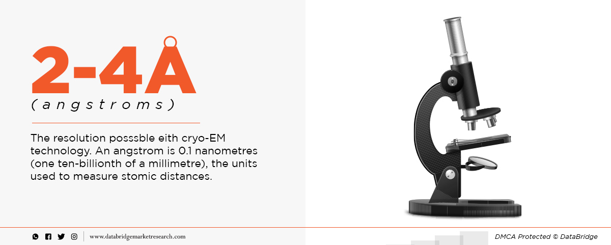
The type of p97 inhibitor binding and contact sites have been observed using Cryo-advanced EM's imaging capabilities. Resolutions of 2.3 ngströms were achieved in this study, with the unit ngström equal to 0.1 nanometers. With current advances in detector technology and sample preparation, cryo-EM can potentially improve further resolution.
Data Bridge Market Research analyses that the cancer diagnostics market is expected to reach the value of USD 28.21 billion by the year 2029, at a CAGR of 7.29% during the forecast period. The rise in the Cancer cases provides growth opportunities to the market. Rising technological advancements is the vital factor escalating the market growth, also increasing initiatives undertaken by governments and global health organizations to spread awareness about cancer, rising growth in the number of private diagnostic centers, rising public-private partnerships to enhance the infrastructure of diagnostic imaging centers, rising FDA support for biomarker development and rising launch of new flow cytometry reagents for diagnostics and drug discovery are the major factors among others driving the cancer diagnostics market.
To know more about the study, visit: https://www.databridgemarketresearch.com/reports/global-cancer-diagnostics-market